MITSUBISHI P91W DRIVER DOWNLOAD

| Uploader: | Kigamuro |
| Date Added: | 12 October 2007 |
| File Size: | 16.82 Mb |
| Operating Systems: | Windows NT/2000/XP/2003/2003/7/8/10 MacOS 10/X |
| Downloads: | 14749 |
| Price: | Free* [*Free Regsitration Required] |
Save enables you to save a protocol or image file once the protocol or image is named. The range of ultraviolet light used by the system. Tile Horizontal places all open image files from top to bottom.
Multichannel image files are saved with an. Use one of the volume tools to create a volume in a representative background region of your image that is, a nondata region similar to the background surrounding your data. Use a flat screwdriver to turn the slotted front of each fuse holder counter clockwise; the holder pops out so you can extract the fuse.
Each new volume you create initially has a red border, which indicates that the volume is selected.
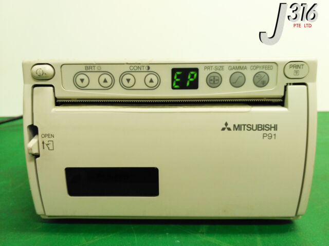
Mitsubishi P91w Digital Monochrome Ultrasound Medical Thermal Printer 13310
In the File menu, select Create Multichannel Image. Regression Method—four regression methods are available.

The R2 value may be used to determine the overall quality of the linear fit. Right-click and select Crop.
Close All closes all the screens. Font—first click the text box you want to change. The primary advantage of this method is that it is extremely simple. To replace a starter, insert it into the holder and rotate clockwise.
MITSUBISHI P91W OPERATION MANUAL Pdf Download.
It is best to use the vertical table orientation when copying to an 8. Position your sample inside the imager and follow the onscreen steps to run a protocol with only one click. Drag a file for each channel into the appropriate channel box in the right pane.
Maximum Value—intensity of the pixel with the maximum intensity inside the volume.
Mitsubishi P91W Manuals
You can zoom in on an area in a current view to export only that area, or you can export the entire image. Configure each channel separately.
The minimum to maximum range varies depending on the light and dark values present in the image. Click and drag the model to rotate it into your preferred view.
Mitsubishi P91W Manuals
To review the relative band quantities: This monitor can be safely placed in patient care areas, such as the operating room, where contamination may occur. No recalibration is necessary to use the gel p1w template kit. Doing so allows valid comparisons between different lanes. Right-click again to return to the original view. CROP You can save crop settings and use them to crop other images.

The calibration need not be changed unless you add equipment, such as a new light source. If you have used colorimetric prestained standards for a chemiluminescent blot, you can acquire an epi-white light image of the blot to show the standards and a chemiluminescent image to show immunodetection. Images should be displayed with consistent, film-like precision and without visible noise or disturbances from the electronic pixel structure.
Refer to page 99 for more information about molecular weight. The electro-magnetic susceptibility has been chosen at a level that gains proper. Instrument Setup enables you to review the instrument serial number and how the imaging system is calibrated.

Comments
Post a Comment